Digital Microscopes
A microscope that can observe uneven surfaces and 3D objects with clear images thanks to its large depth of field and long observation distance. Together with a model that uses a full control system to provide high-resolution observation that rivals an SEM, this lineup includes entry models equipped with frequently used functions. Dedicated lenses that bring out the best performance of the microscopes are also available.
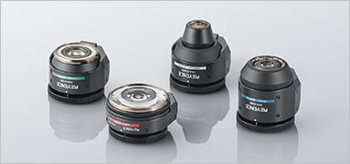
Products Lineup
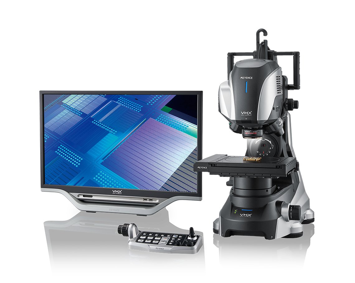
Our flagship model within the VHX Series, new lighting and imaging modes to bring out even the most subtle surface details. With fully-motorised operation, this system supports a wide range of imaging and analysis capabilities including elemental analysis, metallurgical analysis with new high-resolution objectives and software, a 300 mm stage for wafers and other large parts, and functions for image comparisons and automation.


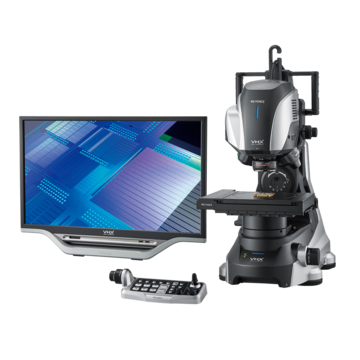
The VHX-XF Series is an all-in-one system for easily viewing, capturing, and measuring parts. This microscope incorporates many of the fundamental features of our VHX-X1 Series in a more budget-friendly package.
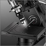
Digital microscopes use a camera and magnified optics to output a live image to a monitor. From the monitor, the image can be observed, captured, saved, measured, and analysed. Unlike traditional optical microscopes, digital microscopes do not have eyepieces, and multiple people can observe the image at the same time.
Benefits of Digital Microscopes
Compared to optical microscopes, digital microscopes have a much larger depth of field.
The depth of field is the z-range that is in focus at one time.
A deep depth of field makes observation easier because the entire sample can be viewed without adjusting the lens or stage. A deep depth of field is especially advantageous when viewing samples with uneven surfaces or large changes in height across the surface.
Digital microscopes offer a larger working distance (WD) than optical microscopes. The working distance is the distance from the end of the lens to the target when the target is in focus.
The longer the working distance, the farther into the target the user can observe. Even when the lens is tilted for observation purposes, the lens does not come into contact with the sample. This is especially useful when working with samples that have large changes in height or have features on different Z planes.
Most optical microscopes use a revolver for switching between the objective lenses to change the magnification.
On the other hand, 3D digital microscopes commonly use a zoom lens that the user merely needs to turn the zoom ring in order to change the lens magnification.
With the revolver, if the working distance differs among the lenses, the user must adjust the stage and focus every time the magnification is changed. With zoom lenses, the working distance remains constant, so users can change magnification while still keeping the sample in focus.
Digital Microscopes Case Studies
Automotive and Aerospace Industries
This section introduces applications such as measurement and analysis of contamination of parts in the automotive and aircraft industries, detection of casting errors such as blowholes, and quantification of wear in cutting tools to manage useful life.
It explains the impact of contamination on automotive and aircraft parts, possible causes of poor die casting and how to counter them, and the mechanism of tool wear. The importance of these inspections and issues that arise with conventional methods are also explained.
Electronic Device Industry
This section introduces an example of failure analysis of implemented PCBs and electronic PCBs carried out by observing the cross-section and surface of semiconductor packages and the wire bonding of electronic components. Typical inspection and observation issues are explained, including potential solutions for capturing clear images and measurements.
Medical Device and Cosmetics Industries
This section introduces applications such as measurement and inspection of medical needles, catheters, stents, pacemakers, and other medical items where high levels of quality are required. Examples of applications in the cosmetics industry, where accuracy and speed are required in the R&D phase, include observation, measurement, and assessment of skin and hair. The current state of the industries is also explained along with case studies of observation using the latest 4K digital microscope.
Chemical and Materials Industries
Welding installations in chemical plants require highly advanced welding quality. The multi-layer films used in food and pharmaceuticals require high levels of safety and reliability. This section introduces observation of penetration of welded parts, as well as examples of different observations and analyses in the chemical and material industries, including high-functionality multi-layer films.
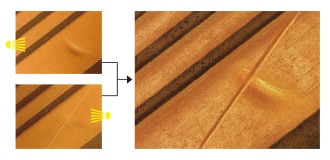
Analysis of Fractured Metal Surfaces
When observing each of the features of a three-dimensional object, such as fracture surfaces, analysis can take a long time due to the need to adjust the focus many times.
The VHX Series 4K Digital Microscope’s real-time depth composition function allows you to focus across an entire fractured metal surface. This allows for observation and assessment of the many composite features that are present in fractured surfaces, in addition to reducing the time spent adjusting the focus.
Observation, Measurement, and Evaluation of Skin and Hair
When evaluating the depth and fineness of surface features, the smaller the height differences in the features, the lower the contrast becomes, which makes observation and assessment difficult. Conventional microscopes show images differently depending on lighting and angle, and are prone to having glare from reflections and lower contrast. This makes shallow grooves difficult to observe, and they can escape detection.
The VHX Series 4K Digital Microscope’s multi-lighting function can automatically capture omnidirectional lighting data. By selecting the image most suitable for observation from this data, you can significantly shorten the time needed to set up the lighting.
Observation of Metal Structures
Observation of metal structures takes time because only a part of the field of view is in focus. Observation and analysis of objects such as this also has a learning curve, which is an obstacle to achieving precise results.
The VHX Series 4K Digital Microscope’s real-time composition interface provides depth composition that allows for quick and easy observation of fully focused images. Additionally, automatic grain size analysis can be performed, eliminating user subjectivity.
Observation and Analysis of Microorganisms
Many microorganisms are highly transparent, which makes it hard to obtain contrast in the observation image. As such, high-magnification observation and quantitative analysis are very difficult for filamentous fungi, which grow in three dimensions, and small bacteria. VHX Digital Microscopes are equipped with multiple different lighting and imaging functions, such as transmitted polarised illumination and HDR, that make it possible to obtain images with high colour gradation. Furthermore, the VHX Series is capable of automatically counting the number of colonies, and providing statistics about each individual colony's size.
Polarised Light Observation for High-Resolution Imaging of Minerals
In polarised light observation of minerals, it is necessary to accurately perceive changes due to the observation method and angle. However, evaluations can differ from one individual to another because it is very difficult to properly set the lighting conditions. The VHX Series supports polarised light observation with parallel or crossed Nicol prisms, and can also perform transmitted polarised illumination. Furthermore, the VHX Series has the ability to recall lighting conditions from previous images, ensuring consistent results.
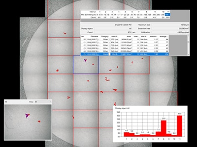
Observation and Measurement of Semiconductor Wafers and IC Designs
VHX Series Digital Microscopes use Optical Shadow Effect Mode, a whole new method of microscopy, to perform high-magnification observation with image clarity that rivals that of an SEM. Optical Shadow Effect Mode enables observation of wafer surface conditions, defective films, and foreign particles. Automatic measurement of photomasking areas and 3D shapes can also be performed. The advanced capabilities of the VHX Series greatly improves inspection and analysis capabilities for wafers and integrated circuits.
Compared to other types of microscopes, digital microscopes are characterised by their high observation functionality and ease of use that is independent of the user’s skill level. However, the required performance is quite different, as is the price of the microscope, depending on whether you are using it for personal observation, quality control in manufacturing, or observation for research. Here, we introduce key points to consider when selecting a digital microscope that is suitable for your intended application.
Magnification and field of view (observation range)
The optical magnification of a stereoscopic microscope can be calculated by multiplying the magnification of the objective lens by that of the eyepiece. In the case of digital microscopes, because images are observed on a screen, the magnification is the product of the lens’ optical magnification and the monitor display size. This magnification is called the total magnification, which can be calculated with the following formula:
Total magnification = Monitor magnification x Optical magnification
The optical magnification is indicated by the lens scale. The monitor magnification differs depending on the image sensor and monitor size, and can be calculated with the following formula:
Monitor magnification = (Monitor size in inches x 16*) / Size of image sensor*
* Optical inch size
Conversely, the size of the field of view (observation range) is inversely proportional to the total magnification. For example, consider a magnification of 50x with a 10 x 6 mm area displayed on the monitor. If the magnification is increased to 100x or 200x, the field of view displayed on the monitor decreases to 5 x 3 mm or 2.5 x 1.5 mm, respectively. As shown in this example, while increasing magnification can enlarge the image for better viewing of details, it also narrows the field of view.
A digital microscope has objective magnification, total magnification, and monitor magnification, which means that when selecting a magnification, you must be aware which of these is indicated in catalogues and which part of the observation object you want to study in an enlarged view. Then, you can select the digital microscope with the magnification and field of view that matches your needs.
Illumination method
When using digital microscopes, you can encounter unclear views of objects despite using a high-performance lens if the lighting is not appropriate. To solve this problem, it is important to choose the type of lighting that suits the object to be observed.
There are four major types of illumination, and this section explains the characteristics and advantages of each type.
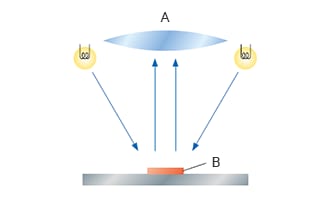
Coaxial illumination
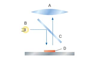
A: Lens, B: Light source, C: Half mirror, D: Object
Coaxial lighting travels in the same direction as the optical path of the lens. With coaxial illumination, the optical axis of the light emitted onto the object and that of the lens are aligned with a half mirror. This mode of illumination is used for observation of mirrored metal surfaces, smooth plastic surfaces, and objects that specularly reflect light, such as semiconductor wafers, as well as when observing differences in structure or surface gloss instead of features.
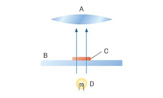
Ring illumination
A: Lens, B: Object
Ring lighting is emitted diagonally from both sides of the lens. This mode of illumination allows for clear capture of contours due to the contrast created by surface features. It is primarily used at magnifications from 50x to 300x for observing objects with rough surfaces or objects with no gloss or lustre.
Transmitted illumination
A: Lens, B: Glass plate, C: Object, D: Light source
With transmitted lighting, light from beneath a transparent target and passes through the lens into the image sensor. For this reason, lenses with a large depth of field are suitable considering the thickness of targets. This mode of illumination is used for observation of features within a transparent object, emulsions in liquids, and microorganisms.
Variable illumination
The direction of light can be changed in real time to emphasise the status of features. Light is emitted onto the target and can be adjusted in real time, allowing for observation of fine features.
Monitor display
One of the most useful features of digital microscopes is that more than one person can look at the microscopic image at the same time. This allows for capturing parts of interest as videos and displaying detailed parts on a monitor for visual checking by multiple people, which can facilitate quick sharing of problems.
The resolution of the monitor can vary from around 2 megapixels to 10 megapixels and up. It is important to select the resolution according to the purpose, along with the selection of magnification and field of view (observation range).
Recording/measurement function
Sharing observation data acquired with a digital microscope between different departments for analysis and review requires functionality that supports large storage capacity and networking. Some key factors to consider include whether observation images can be saved, whether measurement magnification and lighting settings can be saved, and the availability of analysis software that measures profiles, areas, and counts from observation data.
Example: contamination analysis
Characteristics of KEYENCE microscopes
KEYENCE's digital integrated microscopes use a telecentric HR lens, a 4K CMOS image sensor, and a 27-inch 4K monitor. The combination of a high-resolution, large depth of field lens, a high-definition, low-noise image sensor, and a large 4K monitor allows for observation with a wide field of view at high magnifications.
For lighting, KEYENCE’s digital microscopes are equipped with a multi-lighting function that automatically applies the optimal lighting pattern. With just one click of a button, the operator can automatically obtain omnidirectional lighting data and select the image that is most suitable for observation.
Saving on-screen images, measurement results, and observation settings is also only one click of a button away. Saved data can quickly be shared throughout your company via a network connection. Reports can be created by installing spreadsheet and word-processing software.
KEYENCE’s digital microscopes are a one-stop solution that not only eliminate burdensome lighting setups while achieving high-resolution observations and high-quality display, but also provide analytical and reporting functions for data sharing and report submission.
Telecentric HR lens with high NA and high resolution
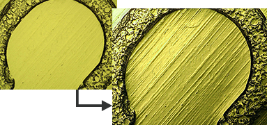
Multi-lighting observation (polished metal surface 1000x)
We have led the way in industrial automation and inspection equipment since 1974. Our commitment to advanced technology and meeting our customers' needs have elevated us to a leadership position in the marketplace. If you're ready to buy digital microscopes from the recognised leader in the field, browse our catalogue and get in touch today.
The basic principles and main types of microscopes are explained, including typical performance, illumination method, and observation method.
Frequently Asked Questions About Digital Microscopes
Digital microscopes are used to observe, inspect, and analyse samples of nearly any size. Digital microscopes are often used as a solution when needs cannot be met by optical microscopes or SEMs. A wide range of industries including electronics, medical devices, materials research, and automotive all use 3D digital microscopes. More information on applications for each industry can be seen on our Application Examples Page.
Digital microscopes from KEYENCE continue to be the best solution on the market. KEYENCE created the first digital microscope over 20 years ago, and continuously implements direct feedback from customers into subsequent generations of scientific digital microscopes. This enables KEYENCE to always be on the bleeding edge, providing products that solve real problems that users face.
Digital microscopes, compound microscopes, stereoscopes, metallurgical microscopes, and polarising microscopes are all commonly used. While the latter four types of microscopes each have a special designated purpose, a digital microscope combines all of these functions into one device, enabling faster throughput and a more efficient workflow.
A digital microscope is equipped with a high-precision digital camera that captures images of the target, which are displayed on the monitor for observation. A stereoscopic microscope has two eyepieces―one for each eye―for observing the image that appears through the lenses. The magnification range of a digital microscope is several tens to thousands times while that of a stereoscopic microscope is only ten to several tens times. A stereoscopic microscope also needs adjustments based on personal differences such as distance between the eyes and visual capability.
Generally speaking, a digital microscope is suitable for long hours of observation, positioning, and dimensional measurement. A digital microscope is also capable of saving images to an HDD as well as image processing and analysis using digital tools. A stereoscopic microscope, on the other hand, has a wider field of view and a longer working distance, and thus it is used for assembling and inspecting precision parts as well as for dissection and cell manipulation.
KEYENCE has been making digital microscopes for over 20 years. We continuously apply the feedback from our customers in the creation of subsequent generations of products. This enables us to always provide products that solve real problems that users face. The digital microscopes in the VHX Series have been designed to compensate for the weak points of conventional optical microscopes such as shallow depth of field, short working distance, problems with portability and versatility, and limitations on samples.